Dr. Martin Johnson joins WoundCentrics San Antonio Wound Clinic  

Martin Johnson, M.D., a Plastic Surgeon and Wound Specialist, has joined WoundCentrics as a wound care provider at the CHRISTUS Santa Rosa Wound Care and Hyperbaric Center at the Medical Center.  He has moved his long time Plastic and Reconstructive Surgery practice from the Phoenix Arizona market to San Antonio.   

An Arizona native that attended Arizona State University, Dr. Johnson received his Medical Degree from Baylor College of Medicine in Houston, Texas.  He completed his Internship, Residency and General Surgery, and a Residency in Plastics Surgery at the Medical College of Virginia, Richmond, Virgina. 

Dr. Johnson is Board Certified by the American Board of Plastic Surgery and is a certified Wound Care Specialist. He recently completed educational credits at Wound Care University in New Braunfels, Texas. 

“We are pleased that Dr. Johnson is bringing his clinical, research and leadership experience to WoundCentrics at the CHRISTUS Santa Rosa Wound Care and Hyperbaric Program in the Medical Center at San Antonio, Texas,” stated Dr. Marcus Gitterle, Chief Medical Officer for WoundCentrics.

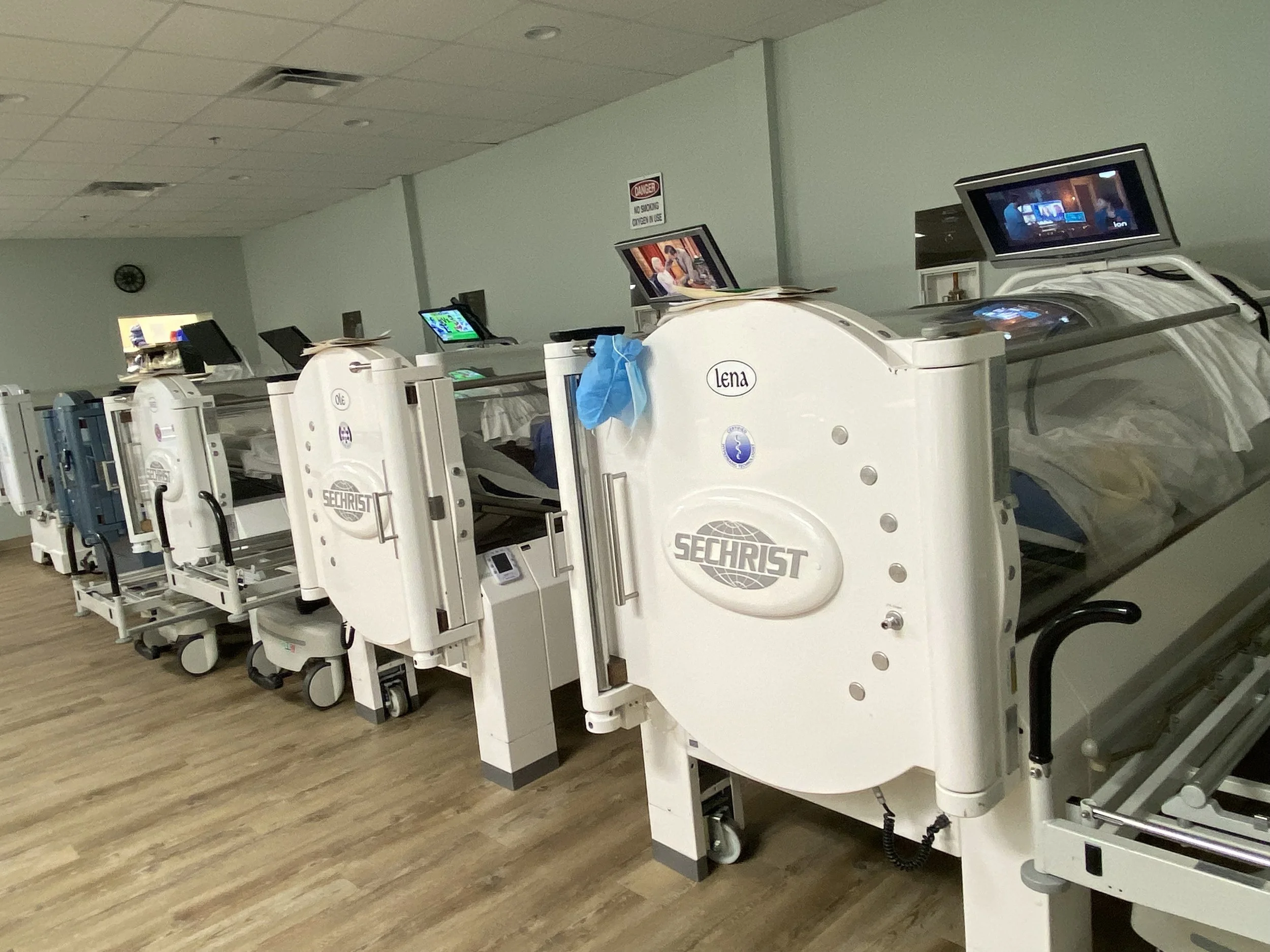
Dr. Johnson will focus on treating patients with chronic, non-healing wounds, including diabetic foot ulcers, arterial and venous ulcers, pressure wounds and bone infections.  Dr. Johnson will oversee the Hyperbaric Oxygen Therapy Treatments in the clinic too.  Dr. Johnson will be accepting new patients at the Christus Santa Rosa Medical Center outpatient Wound & Hyperbaric Clinic located at 2833 Babcock Road, Suite 105, Tower II, San Antonio, Tx 78229.   

For more information visit the website at woundcentrics.com

Vitamin K: A Breakthrough Treatment for Calciphylaxis

Calciphylaxis, a severe condition characterized by calcification of small to medium-sized blood vessels leading to skin necrosis and potential organ failure, and an incredibly high one year mortality rate of uo to 70%, has posed significant management challenges. Recent advances, however, have illuminated a promising treatment modality: vitamin K. Specifically, studies indicate that vitamin K's role in activating the matrix Gla protein (MGP) - an inhibitor of vascular calcification - addresses the core pathophysiology of calciphylaxis.

Traditionally, sodium thiosulphate (STS) has dominated as the primary treatment option, lauded for its calcium-chelating and antioxidant properties. However, its effects on patient survival remain questionable, with many studies showing no marked reduction in mortality. Additionally, adverse side effects like nausea, hypotension, and potential exacerbation of hypercalcemia further limit its applicability.

This brings us to the groundbreaking findings of a recent study. In the words of Dr. Sagar U. Nigwekar and colleagues:

"In a 12-week phase 2 study that enrolled 26 patients with calciphylaxis, Sagar U. Nigwekar, MD, MMSc, of Massachusetts General Hospital in Boston, and colleagues found that patients treated with oral phytonadione, a form of vitamin K, was associated with a significantly lower mortality rate compared with placebo at 12 weeks (0% vs 31%). In addition, phytonadione-treated patients experienced significant decreases in the size of a patient’s largest skin lesion, the combined size of all lesions, and pain intensity compared with placebo.” (1).

The implications of this study are profound. Not only does vitamin K supplementation show promise in reducing lesion size and pain intensity, but its potential to slash mortality rates to zero sets it apart from other treatment modalities like STS.

As the Chief Medical Officer of a Wound Care specialty group involved in treating Calciphylaxis in more than 100 facilities in the US, I am frequently called upon to direct treatment of patients with this condition. Since 2019, our group, based on evidence from the Mass General clinical trial referenced in this article, has been using vitamin K as a principal treatment strategy, with excellent results regarding wound healing and mortality risk. There have been no significant adverse events to date. What is discouraging is the fact that since 2019, our team has yet to encounter a clinician who is familiar with this treatment strategy.

We feel that vitamin K will supplant other treatment methods for Calciphylaxis for several reasons.

1. Targeted Action: Unlike STS, which offers a broader approach to symptom management, vitamin K directly rectifies the vitamin K deficiency that inactivates MGP, a crucial player in the pathophysiology of calciphylaxis.

2. Safety Profile: With an appropriately monitored regimen, vitamin K supplementation exhibits a favorable safety profile, overshadowing the numerous side effects associated with STS.

3. Mortality Reduction: The potential of vitamin K to drastically reduce mortality rates in calciphylaxis patients, as demonstrated in Dr. Nigwekar's study, is a breakthrough in itself.

What is also relevant is that vitamin K is found in two major forms: vitamin K1, or Phylloquinone, found mainly in green, leafy vegetables, and K2, or Menaquinone, found mainly in fermented foods that are uncommon in the US, such as Natto.

Importantly, vitamin K1 is mainly active in the liver, producing blood-clotting proteins. In contrast, vitamin K2 is primarily active outside the liver, which is responsible for activating Matrix GLA protein.

Vitamin K2 is 10 times as potent as vitamin K1 as an activator of Matrix GLA protein while being 1/10 as active in the liver in the synthesis of clotting factors, making it the better choice for the treatment of calciphylaxis because it activates that desired protein, while being less likely to foster a hyper-coagulable state.

In Dr. Nigwekar’s Mass General clinical trial, vitamin K1 was utilized (10mg PO, three times per week). Since most hospital formularies do not contain K2, we typically have utilized the vitamin K1 regimen for inpatients. Upon discharge, I believe it is preferable to transition the patient to vitamin K2, 1mg daily. There are a number of OTC vitamin K2 products, which contain from 90mcg to 1mg of K2.

We recommend choosing a preparation containing at least 600mcg of K2, which can be dosed as two capsules, PO daily. By transitioning to K2, therapy can become more specific in activating the desired Matrix GLA protein and limiting the risk of thrombotic complications. Studies such as Schurgers et al., 2007, have found no association between vitamin K2 intake and increased thrombosis risk (2).

While sodium thiosulphate served its purpose when options were limited, the dawn of a more effective treatment strategy is upon us. Vitamin K, by addressing the root cause of calciphylaxis, promises symptom relief and a profound reduction in mortality risk. It is incumbent on those involved in treating this disease to become familiar with vitamin K treatment for Calciphylaxis to reduce the morbidity and mortality risk of this condition.

1. Nigwekar SU, Krinsky S, Thadani RI, et al. Phase 2 trial of phytonadione in calciphylaxis. Presented at the American Society of Nephrology’s Kidney Week 2019 meeting held Nov. 5 to 10 in Washington, DC. Poster TH-PO1188.

2. Schurgers, L. J., & Vermeer, C. (2007). Determination of phylloquinone and menaquinones in food. Effect of food matrix on circulating vitamin K concentrations. Haemostasis, 30(6), 298-307

The Difference WoundCentrics Makes in Specialized Wound Care

In healthcare, wound care often gets less attention than acute and chronic diseases, yet it plays a critical role in improving patients' quality of life. Non-healing wounds, pressure ulcers, and diabetic foot ulcers can lead to severe complications if not effectively managed. That is where WoundCentrics steps in, making a significant difference in caring for patients with complex wounds. Through its specialized providers, trained extensively by Wound Care University, WoundCentrics is redefining what quality wound care looks like. 

At the heart of WoundCentrics' approach is its commitment to excellence in training. WoundCentrics invests heavily in the education of its healthcare providers, offering training programs through Wound Care University. This university is designed to deliver in-depth knowledge and skills specifically for wound care management. Through its specialized curriculum, Wound Care University covers everything from the basic principles of wound healing to the latest advancements in wound treatment modalities. 

The result? Every provider within the WoundCentrics network is well-versed in standard wound care protocols and equipped to handle complex, difficult-to-treat wounds that require a high level of expertise. This ensures patients receive care from professionals specialized in wound management rather than general practitioners with limited wound care training. 

One of WoundCentrics' key differentiators is its dedication to evidence-based care. The knowledge imparted by Wound Care University is rooted in the latest research and medical advancements in wound care. Providers learn about innovative treatments such as bioengineered skin substitutes, negative pressure wound therapy, and advanced wound dressings that can accelerate healing and reduce complications. 

By applying these cutting-edge techniques, WoundCentrics specialists can often achieve better outcomes in less time, reducing the risk of infection and the need for more invasive interventions like amputations. For patients suffering from chronic wounds, this can be life-changing. 

WoundCentrics emphasizes the importance of collaboration in wound care. Providers trained by Wound Care University are taught to work closely with other healthcare professionals, including primary care physicians, vascular specialists, podiatrists, and physical therapists. This team-based approach ensures that the wound care process considers every aspect of the patient's health. 

For example, a non-healing leg ulcer patient may benefit from advanced wound care and a consultation with a vascular surgeon to assess circulation issues. By coordinating care across disciplines, WoundCentrics ensures patients receive the comprehensive treatment they need to heal. 

WoundCentrics sets the standard for wound care by focusing on specialized education, evidence-based treatments, and holistic patient care. Through its Wound Care University, it ensures that every provider is highly trained in the most advanced wound care techniques and equipped to deliver superior outcomes. For patients dealing with the challenges of chronic or non-healing wounds, the difference that WoundCentrics makes is profound: faster healing, fewer complications, and a better quality of life. 

As the demand for specialized wound care continues to grow, WoundCentrics stands out as a leader in the field, delivering expert care that transforms patients' lives. 

Kent Imaging and WoundCentrics Propel Wound Care Forward With New Partnership

WoundCentrics includes SnapshotNIR as part of a system-based approach to patient-centered wound care.

CALGARY, ALBERTA, CANADA, April 2, 2024 /EINPresswire.com/ -- Kent Imaging is excited to be a part of the next steps WoundCentrics is taking to advance its patient care by incorporating SnapshotNIR as a point-of-care imaging tool. WoundCentrics’ system-based approach to each patient prioritizes the rapid acquisition of key data relevant to the patient’s assessment and treatment.

Vascular evaluation is integral to optimal decision-making in wound care. By enabling better vascular assessment at the point-of-care by including SnapshotNIR on initial and follow-up visits, the WoundCentrics team is better able to create optimal patient-centric care plans and achieve better outcomes.

“Our clinicians are trained in the multifaceted nature of wound care decision-making,” states Dr. Marcus Gitterle, MD, Chief Medical Officer of WoundCentrics. He adds, “We envision SnapshotNIR as a key component of risk reduction by having documentation and data from the device to better enable our teams’ clinical decisions.”

Not only does this partnering between Kent Imaging and WoundCentrics benefit patients, but physicians will be able to expand their research capabilities. With Kent’s eye for innovation and WoundCentrics’ mission to continue to contribute high quality research to move the industry forward, this is an exciting opportunity to elevate the standard of wound care.

WoundCentrics is a multi-state wound care provider, servicing patients through multidisciplinary teams with exceptional wound care expertise across all patient care domains including inpatient STACH, LTACH, IRF, SNF, LTC, and the home setting. Their system-based approach includes head-to-toe assessments and re-evaluations of patients through daily care from inpatient wound care providers to weekly outpatient visits in the clinic setting.

“Regardless of the patient care domain, we expect SnapshotNIR to become a key tool in our system-based patient management protocols, providing rapid, actionable, objective data to confirm the clinician’s observation and current diagnostics,” states Dr. Gitterle, he continued, “Faster delivery of this data means more efficient and effective patient care, ultimately giving time back to the patient in circumstances where time can mean saving a limb or making a decision regarding invasive surgical interventions.”

SnapshotNIR is an imaging device that uses reflectance-based technology to measure the hemoglobin and oxygen saturation of tissues 2-3 mm below the skin. Tissue oxygen saturation (StO2) is essential to wound healing and understanding a patient’s health status. Snapshot is a non-invasive, portable device that clinicians can use during bedside assessments, providing instantaneous data to support treatment planning, validate efficacy, and facilitate patient adherence to treatments like hyperbaric oxygen therapy (HBOT), a service WoundCentrics provides.

“We believe in the difference that SnapshotNIR can make in a patient’s healing progression,” says Kent’s Chief Executive Officer, Pierre Lemire, “With Snapshot in the skilled hands of WoundCentrics’ providers, we’re moving in the right direction keeping patients at the center of our cause, improving outcomes in as many patients and places as possible.”

Imaging procedures with SnapshotNIR have applicable CPT codes for reimbursement. To find out more, visit our dedicated reimbursement page online.

About Kent Imaging
Kent Imaging, located in Calgary, Alberta, Canada, is a leading innovator in near-infrared tissue oxygenation imaging, which develops, manufactures, and markets medical technology that supports real-time decision-making in wound care, vascular and surgical subspecialties. Kent holds multiple patents in oxygen imaging technology and continues to provide innovative and advanced diagnostic imaging solutions to aid healthcare systems nationally and internationally. SnapshotNIR is supported by clinical evidence demonstrating its ability to help improve clinical decision-making in wound care and reduce healing time. Since receiving FDA and Health Canada clearance in 2017, the technology has been featured in several published articles and peer-reviewed posters. Applying the knowledge gained from clinical trials to patient care promotes consistency of treatment and optimal outcomes.

About WoundCentrics
WoundCentrics is based in New Braunfels, Texas, USA, and operates fourteen wound centers and provides advanced wound care services in more than one hundred inpatient facilities across twelve states with a goal of providing advanced wound care, amputation prevention and vascular services to these communities.

Leah Pavlick
Kent Imaging Inc.

NEWS PROVIDED BY

Kent Imaging Inc.

April 02, 2024, 13:00 GMT

Platelet Rich Plasma: Expanding Options for Non-Healing, Chronic Wounds

Platelet Rich Plasma: Expanding Options for Non-Healing, Chronic Wounds

A substantial percentage of non-healing, chronic wounds fail to heal, even under expert care by wound specialist providers, necessitating the application of special care, involving “Advanced Modalities,” such as Hyperbaric Oxygen Therapy, and Engineered Dermal Substitutes, sometimes referred to as “synthetic skin.” While such “Advanced Modalities” can provide significant benefits to patients, they are expensive, not universally available, and reimbursement for their use is quite restrictive.

At present, third-party payers restrict reimbursement for engineered grafts to specific wound types and locations; namely Diabetic Foot Ulcers, and Venous Leg Ulcers, and restrict Hyperbaric Oxygen Therapy, as a chronic wound treatment modality to Diabetic Foot Ulcers, and chronic, radiation-related wounds.

Those site and etiology-based restrictions leave many patients with diabetes who also have non-healing ulcers of sites other than the foot, bereft of options. Fortunately, an exciting new option is available for diabetic patients with non-healing ulcers at any site, as long as non-healing is related to underlying complications of diabetes, such as microvascular disease, white blood cell, or fibroblast dysfunction.

The technology we are referring to is Platelet Rich Plasma or PRP, which is not a “new technology,” per se. In fact, PRP has a proven track record in regenerative medicine, and particularly within orthopedic medicine, where it has been used for more than a decade to improve outcomes in patients with complex tendon, muscle, bone and joint problems. In fact, the very basis for the successful application of PRP to orthopedic problems underlies its benefits as an Advanced Modality in wound care.

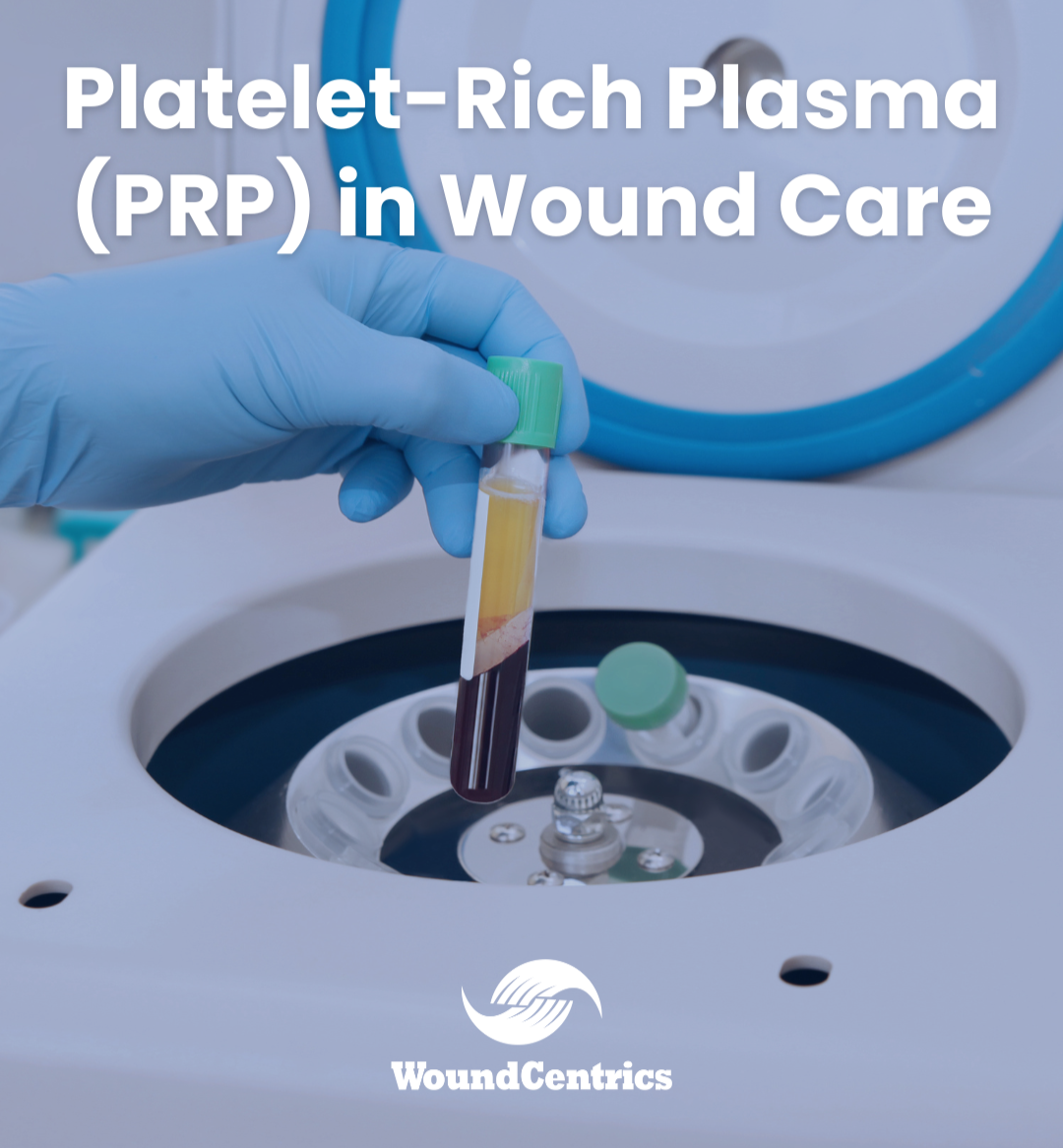
From a technical standpoint, PRP is a biologically derived product — a type of Autograft —  sourced from the patient's own blood. Using a simple process that can be quickly performed in the wound clinic, the patient’s blood is utilized to create a jelly-like graft material that is enriched with growth factors, activated platelets, fibrin, and other bioactive elements essential for tissue repair and regeneration.

Not only does PRP provide a potential solution for diabetic patients without other reimbursable treatment options, it provides a unique combination of benefits that are not seen with existing solutions, such as amniotic grafts. These benefits include:

1. Tissue Regeneration: PRP accelerates the body's natural healing processes via endogenous, patient-derived cytokines, promoting the regeneration of damaged tissue by enhancing synthesis of new blood vessels, and collagen synthesis, both of which are necessary prerequisites for wound healing.

2. Infection Control: PRP exhibits inherent antimicrobial properties, which contribute to infection control within the wound environment, creating a conducive setting for healing, and offsetting some of the detriments to wound healing posed by biofilms — bacterial structures that are found in most, chronic wounds that strongly inhibit healing.

3. Inflammation Modulation: PRP also plays a role in modulating the inflammatory response, a pivotal factor in the pathophysiology of chronic wounds, thus facilitating the healing process.

4. Scar Reduction: Notably, PRP therapy has demonstrated the potential to minimize scarring and improve cosmetic outcomes, a welcome benefit for patients concerned about aesthetic consequences.

The PRP Treatment Protocol is straightforward, and easily repeatable by wound clinic personnel after brief training, and consists of the following, simple steps:

1. Blood Collection: A modest volume of the patient's blood is drawn, mirroring the process employed for standard blood test specimen collection.

2. Centrifugation: The blood sample undergoes centrifugation to isolate PRP from other blood constituents.

3. Application: The concentrated PRP is applied directly to the wound site, and can also be applied via targeted injection, depending on wound characteristics and anatomical considerations. The jelly-like consistency of PRP lends itself to application to complex wound shapes, and sinus tract wounds that would be difficult to cover with planar or sheet materials.

4. Dressing and Monitoring: Following PRP application, an appropriate wound dressing is applied, and close monitoring is conducted to assess the progress of wound healing.

Conclusion

In Platelet Rich Plasma (PRP) therapy, we have a noteworthy addition to our arsenal of wound care strategies. It offers a hope for a wider range of individuals grappling with non-healing, chronic wounds. By harnessing the body's inherent regenerative capabilities, PRP promises expedited wound closure, a reduction in complications, and an improvement in overall quality of life. As we navigate this therapeutic landscape, we stand at the precipice of innovation, holding the potential to significantly enhance patient outcomes and usher in a new era in the domain of wound management. Embracing this emerging approach represents an opportunity for substantive improvements in the treatment of chronic wounds and a reconfiguration of established paradigms in the field.


Marcus Gitterle, MD, FACCWS, ABWM
Chief Medical Officer
WoundCentrics, LLC

Diabetic Foot Care for Patients

If you have diabetes, here’s a way to keep standing on your own two feet: check them every day—even if they feel fine—and see your doctor if you have a cut or blister that won’t heal.  

There’s a lot to manage if you have diabetes: checking your blood glucose, making healthy food, finding time to be active, taking medicines, and going to doctor’s appointments. With all that, your feet might be the last thing on your mind. But daily care is one of the best ways to prevent foot complications.  

There’s a lot to manage if you have diabetes!  

Some people with nerve damage have numbness, tingling, or pain, but others have no symptoms. Nerve damage can also lower your ability to feel pain, heat, or cold. 

What’s the most important thing you can do to prevent nerve damage or stop it from getting worse? Keep your blood sugar in your target range as much as possible. Other good diabetes management habits can help, too: Stop smoking! Smoking reduces blood flow to the feet.  

Follow a nutrition plan, including eating more low-glycemic fruits, such as berries and vegetables, and avoid anything with sugar in it, including fruit juices, sodas, pastries, and candies. Physical activity—10 to 20 minutes a day is better than an hour once a week.  

Take medicine as prescribed by your doctor. Anyone with diabetes can develop nerve damage, but these factors increase your risk:  Blood sugar levels that are hard to manage, having diabetes for a long time, especially if your blood sugar is often higher than your target levels, being overweight, being older than 40 years, having high blood pressure and having high cholesterol.  

Nerve damage, along with poor blood flow—another diabetes complication—puts you at risk for developing a foot ulcer (a sore or wound) that could get infected and not heal well. If an infection doesn’t get better with treatment, your toe, foot, or part of your leg may need to be amputated (removed by surgery) to prevent the infection from spreading and to save your life. When you check your feet daily, you can catch problems early and treat them immediately. Early treatment greatly reduces your risk of amputation.  

One of the best ways to prevent foot infections and amputations is to Inspect your feet daily!  

Get to the bottom of any foot problems by using a mirror or asking for help. Check your feet daily if you have lessened sensation.  

Career Growth & Advancement are A Reality at WoundCentrics

Shellie Torres, Director of Human Resources 

Shellie Torres is the Director of Human Resources for WoundCentrics, a Texas-based Wound Care company. WoundCentrics is a growing healthcare company that is hiring new team members. The current team is creating new opportunities through client growth, which also creates career growth opportunities for existing and new team members. Shellie's team is responsible for the hiring process at WoundCentrics, and she is an example of what career growth can look like in the company.  

Shellie began her career with WoundCentrics in February 2016 as a recruiter. After several job advancements where she took on progressive responsibility, she was ultimately promoted to Director of Human Resources in January 2022.   

In 2017, Shellie decided to grow her career by furthering her education and taking advantage of company benefits to support her career growth. Shellie outlined a plan with her managers at WoundCentrics to achieve the education and career development she received from her involvement in the Lubbock chapter of the Society for Human Resource Management (SHRM) where she eventually became chapter President.  

SHRM is a professional human resources membership association that provides education, certification, and networking to its members, while advocating on issues pertinent to labor management. 

"I was ready for another stage in my career in 2017," stated Torres. "I wanted a new challenge, I wanted to make a difference for our associates, and I knew I needed more education to get there.”  

"I decided to grow within the company, which meant focusing on my education. I completed my bachelor's degree in 2020 and my master's degrees in 2022 which resulted in advancing to a new role and taking on more responsibility,” continued Torres. "I also gained new skills, met new people, and gained a new perspective on my role in the company as the Director of HR. Professional growth and advancement can make you feel more satisfied with your position and more confident in your job-related skills," stressed Torres.  

In today's healthcare market — the more skilled and educated employees are, the better a business performs in operations, outcomes, and profitability. As Shellie's experience shows, employee education and job skills open doors. WoundCentrics is an advocate for and supportive of motivated employees' goals and plans for career growth.  

“I was fortunate to have employers throughout my career that supported career growth through education and professional development, whether that be financially or through time to serve on committees and boards that broadened my knowledge and understanding of my profession and those I worked with. It is an honor and a pleasure to extend that opportunity those who work for WoundCentrics and our family of companies so that they can hopefully have a similar experience,” share Stuart Oertli, WoundCentrics’ Chief Operating Officer.  

A recent Indeed article pointed out that companies benefit from employees who seek out opportunity for career growth. The report said that aside from company perks and monetary compensation, today's top talent looks for employers that enable them to learn and grow professionally.   

As WoundCentrics seeks highly qualified talent in today's healthcare market and as competition for employees continues to increase, the opportunity for career growth is one of the most critical concerns to the modern job seeker when evaluating a prospective employer – and this is also true for existing employees.  WoundCentrics has taken steps to develop in house continuing education, has monthly provider education meetings for it’s direct care providers, has developed provider onboarding for new wound care practitioners and actively encourages associates to attend and present at professional conferences. 

Shellie Torres has talked and walked the talk and is an example of what hard work, commitment, education, and a career plan can yield at WoundCentrics.  

For more about careers at WoundCentrics, visit our careers page.

Debridement: How can we help a wound heal by making it larger?

It is often a point of confusion for patients, family members, and even clinical professionals, when debridement is recommended for the treatment of an acute, or chronic wound. “Why would making a wound larger be helpful, when our goal is to make it heal?"

Debridement is the removal of dead, damaged, or infected tissue from a wound, and it is a standard part of wound care for many types of wounds. The benefits of debridement are well-established and supported by a large body of clinical evidence.

Debridement helps to promote wound healing by removing debris, bacteria, and other harmful substances from the wound bed, which can impede the healing process. It also helps to stimulate the growth of healthy new tissue, which can speed up the healing process.

Several studies have demonstrated the benefits of debridement for wound healing. For example:

  1. A systematic review and meta-analysis published in the Journal of Wound Care in 2019 found that debridement was associated with a significant reduction in wound healing time and a higher rate of complete wound healing compared to non-debridement treatments.

  2. A randomized controlled trial published in the Annals of Emergency Medicine in 2015 found that debridement of traumatic wounds reduced the risk of infection and improved wound healing outcomes compared to non-debridement treatment.

  3. A systematic review and meta-analysis published in the Cochrane Database of Systematic Reviews in 2016 found that debridement was associated with a faster rate of healing and a reduced risk of infection in diabetic foot ulcers.

Overall, the clinical evidence strongly supports the use of debridement as a standard part of wound care for many types of wounds. It's important to consult with a healthcare provider to determine the most appropriate debridement technique for a specific wound, as there are different types of debridement, including surgical, mechanical, enzymatic, and autolytic debridement, and the most appropriate technique may depend on the type and severity of the wound.

WoundCentrics providers receive extensive training in the theory and techniques of debridement, making them uniquely capable of delivering high-quality wound care, tailored to the Whole Patient, not just the hole in the patient.

Omega 3 Fats are Finding a New Role in Wound Therapeutics

Omega 3 Fats are Finding a New Role in Wound Therapeutics

Omega 3 Fatty acids are well known to most people these days as an important component of a healthy diet. They are known to impact heart health, and reduce inflammation, but, could they be useful in wound healing?

Yes, there is research supporting the use of topical omega-3 fatty acids to treat wounds. Omega-3 fatty acids have been shown to have anti-inflammatory and wound healing properties, making them potentially useful in the treatment of various types of wounds.

A study published in the Journal of Investigative Dermatology in 2013 found that topical application of an omega-3 emulsion helped to accelerate wound healing in mice. The study found that the omega-3 emulsion reduced inflammation and increased collagen synthesis, which are important factors in the wound healing process.

Another study published in the Journal of Wound Care in 2018 looked at the effects of a topical omega-3 dressing on the healing of diabetic foot ulcers. The study found that the omega-3 dressing was associated with faster wound healing and a reduced risk of infection compared to a standard dressing.

Overall, while more research is needed to fully understand the potential benefits of topical omega-3 fatty acids in wound healing, there is evidence to suggest that they may be a useful addition to standard wound care treatments. It's important to consult with a healthcare provider before using any new treatments for wounds or other medical conditions.

At WoundCentrics, we will be following Omega 3-based topical therapies, as well as scanning the horizon for breakthroughs that can help our provider team heal our patients as fast as possible. Staying abreast of the latest developments in the science of wound healing is part of our mission.


Engineered Dermal Substitute

Chronic wounds are a growing challenge for medical professionals, and systems. While there are fundamental interventions that are applicable to most or all problem, or chronic wounds, such as wound bed preparation, supplemental nutrition, optimization of co-morbid conditions such as diabetes, and assessment and correction of vascular deficits, if present, no all wounds heal, even when best-practices are followed, and these areas addressed.


In the effort to facilitate healing in patients who fail to respond to full-spectrum standard wound care, advanced modalities are an increasingly important option. One type of Advanced Modality is called an Engineered Dermal Substitute. There are now more than 700 FDA approved dermal substitute products, and several distinct sub-categories within this larger grouping.


An engineered dermal substitute (EDS) is a type of skin substitute that is used to treat wounds or defects in the skin. It is made of biocompatible materials that mimic the structure and function of human skin, and is designed to promote the growth of new tissue.


EDS typically consists of a three-dimensional scaffold made of biocompatible materials such as collagen, hyaluronic acid, or synthetic polymers. This scaffold is seeded with cells such as fibroblasts or keratinocytes, which are key cells involved in the healing process. The cells grow and organize within the scaffold, creating a new layer of tissue that can integrate with the surrounding skin.


EDS can be used to treat a variety of skin injuries, such as burns, chronic wounds, and ulcers. It can also be used for reconstructive surgery to replace lost or damaged skin. EDS has several advantages over traditional skin grafts, including a reduced risk of infection, less scarring, and the ability to customize the scaffold to fit the specific needs of the patient.


While EDSs have shown promise in clinical trials, they are still a relatively new technology and further research is needed to determine their long-term safety and effectiveness. However, they have the potential to be an important tool in the treatment of chronic wounds, and could help improve the quality of life for patients with these conditions.

The Effects of Smoking on Wound Healing

A chronic wound is not an isolated problem, but is often a symptom of chronic diseases that occur due to genetics and lifestyle choices. Although genetic makeup is not a modifiable factor, lifestyle choices are. Specifically, tobacco use is a choice that can significantly impair wound healing. However, there is good news for those patients who use tobacco products and suffer from chronic wounds. By giving up tobacco use, one can significantly improve wound healing and reduce overall wound care expenses.  

American culture tends to emphasize smoking cessation and the dangers of tobacco use in public school education and medical practices, yet according to the CDC (2022) about 30.8 million American adults still pursue habitual tobacco use. Furthermore, smoke exposure through second-hand smoke reaches a broader range of Americans with 41,000 deaths annually among non-smoking adults (CDC, 2022).  

Smoking increases overall mortality risk and negative health outcomes. Specifically, with wound care, the effects can be detrimental to wound healing. Smoking negatively impacts wound healing by impairing the immune system and reducing blood flow in the skin required for healing. Blood circulation delivers oxygen and nutrients to the cells, promoting the closure of wounds. Nicotine causes vasoconstriction which is the narrowing of blood vessels. Vasoconstriction reduces the perfusion of the wound bed and can delay tissue growth, thus delaying healing.  

The immune system is also impaired with nicotine use because white blood cells depend on normal oxygen levels in order to kill bacteria. By lowering tissue oxygen levels increasing risk of infection, which can lead to abscess formation, wound treatment failure, limb amputation, and even premature mortality. Delivery of nutrients to the wound bed is also decreased with tobacco. Nutrients like Vitamin C are a vital component needed in wound healing along with Vitamins A, D, and zinc.  

Through nicotine exposure, whether from cigarette smoking, vaping, smokeless tobacco, or even nicotine gum, the overall process of wound healing is hindered, thus increasing the physical and financial demands on the patient. Delayed wound healing can increase doctor visit frequency, loss of wages, use of wound care products, prescriptions, and financial burden needed to address the open wound. The risk of continuing tobacco use can be detrimental to overall health.  

The great news is that a 2016 study showed patients who stopped smoking for eight hours had oxygen levels normalize in the bloodstream. The same study showed that after 24 hours of smoking cessation, nerve endings started to recover. After two weeks, blood flow improved and helped increase mobility. The dangers of smoking to wound healing and overall health far outweigh the challenges of smoking cessation. Resources are available to assist with smoking cessation. Chronic wounds are challenging to heal, but smoking cessation can expedite the healing process. 

Proper Nutrition Enhances Proper Wound Healing

Proper Nutrition Enhances Proper Wound Healing

A significant number of people who have acute, slow healing, or chronic wounds are nutritionally impaired.  Proper wound healing is enhanced by taking in adequate amounts of dietary protein, Vitamin A, Vitamin C, Vitamin D, and Zinc, as well as hydration.  Many people who have wounds eat a diet that consists of heavy amounts of carbohydrates and fats and not enough proteins, fruits, and fresh vegetables.   

Wound healing occurs in 4 distinct phases.   

  • Hemostasis- This phase occurs immediately after the wound is created and refers to a series of biologic processes which result in decreased bleeding.  

  • Inflammatory Phase- This phase starts as bleeding is being controlled. During this phase a variety of specialized white blood cells and other immune system mediators are transported to the site of injury. This phase usually lasts for 2 weeks and causes localized swelling, pain, tenderness, and redness.  

  • Proliferative phase- This phase usually lasts for 3 to 4 weeks. In this phase large numbers of special cells called fibroblasts produce collagen. Collagen is a protein which is the primary “building block” of the structure of all live animals. Without collagen there would be no bones, muscle, skin or wound healing. This is the phase in which the wound becomes smaller by contraction and is covered with new skin. This is also the most common phase in which wounds “get stuck” and stop healing.  

  • Remodeling- This is the final stage of wound healing and lasts for up to 1 year after the wound was formed. During this time collagen fibers become linked together and increase the strength of the final scar. The scar continues to shrink and changes color from red to the normal skin color. 

Proper nutrition is necessary for the phases of wound healing to proceed normally. The single most important nutritional supplement needed is protein. Dietary protein is broken down into amino acids which are used by fibroblasts to synthesize the new structural proteins and are essential for wound healing and the repair of injured tissues.  If dietary intake of protein is inadequate, healthy protein from other body tissues will be broken down and used for wound healing. This is obviously counterproductive for the otherwise healthy tissues such as muscle and bone. It is recommended to take protein supplements in addition to animal protein eaten daily with meals. There are numerous products available at most grocery stores and pharmacies. The recommended dose is 2 servings per day of a 30gm protein supplement. Patients with diabetes need a supplement that has reduced sugars, and patients with renal failure or liver problems may also require a specialized protein supplement to avoid metabolic complications.  Please consult with your physician if you have renal failure or liver problems prior to purchasing an over the counter supplement. 

Several specific vitamins and minerals are also crucial for the phases of wound healing to proceed normally. 

Vitamins A, C, D, and Zinc are required in various stages of the wound healing process and in the regulation of a healthy immune system as well. Normal daily multivitamins do not contain adequate amounts of these to enhance wound healing. In addition to a daily over-the-counter vitamin the recommended doses of these are as follows:  

  • Vitamin A- 5000 units per day 

  • Vitamin C- 2000mg per day 

  • Vitamin D3- 5000 units per day 

  • Zinc- 50mg per day 

In summary, proper nutrition is one of key factors in proper wound healing. The best results in successful healing of difficult wounds are seen in a qualified Wound Care Center where the providers are trained in taking care of the whole patient to ensure wound healing.  They review important factors involved in wound healing and monitor each patient closely until a successful outcome is achieved. 

Wound Care University to train all WoundCentrics Physicians, Advanced Practice Providers and Allied Health Professionals on Hyperbaric Medicine and Advanced Wound Care

WoundCentrics, a full-service wound care management company, uses Wound Care University in New Braunfels, Texas exclusively to train its employed and contracted physicians, advanced practice providers and allied health professionals for hyperbaric medicine training. The Wound Care University 40-hour Introduction to Hyperbaric Medicine course is an Undersea Hyperbaric Medicine Society approved course and offers CME to the physicians that complete the course and pass the exam.

“Our company offers all our providers the clinical training needed to achieve their advanced wound care and hyperbaric medicine certifications. Wound Care University provides the quality education and professional advancement that our company is seeking to offer our clinicians,” stated Stuart Oertli, Chief Operating Officer of Wound Centrics.

“The basic and advanced wound care training provided by Wound Care University builds the foundation needed for the WoundCentrics clinical providers to become proficient at the various therapies, modalities and treatments required to be an advanced wound care professional in our industry,” according to Marcus Gitterle M.D, Chief Medical Officer of WoundCentrics.

“We train our providers in advanced wound care to be able to work in the acute hospital inpatient setting, and in the post-acute setting, including the rehabilitation hospitals, long term acute care hospitals, skilled nursing home facilities, and long-term care,” Oertli explained. “The expanding sub-specialty of wound care is evolving rapidly, and our providers are trained to be up to date and proficient on the latest evidenced-based treatments of wound healing,” explained Dr. Gitterle.

WoundCentrics was founded 12 years ago in San Antonio, Texas. The company has grown to have a large national footprint, operating in ten states and in more than ninety-two facilities. Through its physician employment group, Physician’s Unity, it employs sixty-four providers and more than seventy-five other clinical, administrative, and support staff.

In 2019, Wound Care University was founded to provide state-of-the-art wound care training and development for wound care clinicians, including an Undersea and Hyperbaric Medical Society (UHMS) approved course for new hyperbaric clinicians (www.woundcareuniversity.com). To learn more about WoundCentrics visit the website at www.woundcentrics.com.

WoundCentrics Supports Focus on Diabetes Awareness and Education

WoundCentrics Supports Focus on Diabetes Awareness and Education

Diabetic Foot Ulcer.jpg

November is American Diabetes Month

WoundCentrics is honoring Diabetes Month by joining the awareness and education campaign in the communities where we provide specialized wound care services. Currently, 34.2 million Americans have diabetes and 2 million are struggling to heal a diabetic foot ulcer. America’s diabetic population is expected to nearly double by 2030, it is important to know the risks associated with diabetic foot ulcers according to the American Diabetes Association:

  • Up to 25% of people living with diabetes will experience a foot ulcer in their lifetime

  • 14-24% of foot ulcers progress to amputation

  • 85% of diabetes-related amputation were preceded by a foot ulcer

  • 130,000 for a lower-extremity amputation (5.6 per 1,000 adults with diabetes)

  • 50% of patients die within five years of amputation

WoundCentrics provides advanced wound care services that identify and specializes in treating wounds, including diabetic foot wounds.  WoundCentrics wound clinics coordinate wound care services with other healthcare services treating diabetic patients with other diabetic complications, manages and heals wounds, prevents amputations, and preserves the quality of life for diabetic patients.

WoundCentrics Wound and Hyperbaric Centers and the WoundCentrics Providers are specially trained in wound care and diabetic foot ulcer care, including hyperbaric oxygen therapy, a treatment for diabetic ulcer patients.

WoundCentrics’ mission is to support the National Diabetes Month to increase awareness of the risks, along with proper care for diabetic foot ulcers that can reduce diabetes-related amputations in the communities that they offer wound care services. For more information on WoundCentrics visit www.woundcentrics.com

Diabetes and the Complication of Diabetic Foot Wounds

According to the National Institute of Diabetes and Digestive and Kidney Disease (NIDDK), a division of the National Institute of Health (NIH),  National Diabetes Week brings awareness and focus to the major health issue of diabetes and the complications of diabetes like diabetic foot wounds and diabetic neuropathy.

Foot problems are common in people with diabetes. You might be afraid you’ll lose a toe, foot, or leg to diabetes, or know someone who has, but you can lower your chances of having diabetes-related foot problems by taking care of your feet every day. Managing your blood glucose levels, also called blood sugar, can also help keep your feet healthy.

WoundCentrics manages patients suffering from diabetes and foot complications and wounds every day.  If you have diabetes and a foot wound, WoundCentrics specialist and our wound and hyperbaric centers can help heal your wound and return patients to a better quality of life.

For more information on diabetes and foot care from the National Institute of Health visit https://www.nih.gov/

Diabetes Awareness Month

American Diabetes Awareness Month (or simply Diabetes Month) is an annual campaign throughout the month of November in the USA to bring awareness to the growing prevalence of diabetes, the health risks associated with it, raise funds for research into the condition, and support people living with it.

Diabetes is one of the fastest-growing, preventable medical conditions in the world.

Recent research by the Diabetes Research Institute published in 2020 also points to some alarming statistics. Among the US population overall the prevalence of diagnosed and undiagnosed people with the condition for 2018 were (crude estimates):

  • 34.2 million people of all ages—or 10.5% of the US population—had diabetes

  • 34.1 million adults aged 18 years or older—or 13.0% of all US adults—had diabetes

  • 7.3 million adults aged 18 years or older who met laboratory criteria for diabetes were not aware of or did not report having diabetes (undiagnosed diabetes). This number represents 2.8% of all US adults and 21.4% of all US adults with diabetes

  • The percentage of adults with diabetes increased with age, reaching 26.8% among those aged 65 years or older

Diabetes also affects different ethnic groups differently. The prevalence of diagnosed diabetes was highest among American Indians/Alaska Natives (14.7%), people of Hispanic origin (12.5%), and non-Hispanic blacks (11.7%), followed by non-Hispanic Asians (9.2%) and non-Hispanic whites (7.5%).

The condition also causes many deaths:

  • In 2017, diabetes was the seventh leading cause of death in the United States. This finding is based on 83,564 death certificates in which diabetes was listed as the underlying cause of death (crude rate, 25.7 per 100,000 persons)

  • In 2017, there were 270,702 death certificates with diabetes listed as the underlying or contributing cause of death (crude rate, 83.1 per 100,000 persons)

And the costs are astronomical!

  • The total direct and indirect estimated costs of diagnosed diabetes in the United States in 2017 was $327 billion

  • Total direct estimated costs of diagnosed diabetes increased from $188 billion in 2012 to $237 billion in 2017 (2017 dollars); total indirect costs increased from $73 billion to $90 billion in the same period (2017 dollars)

  • Between 2012 and 2017, excess medical costs per person associated with diabetes increased from $8,417 to $9,601 (2017 dollars)

For the individual, the American Diabetes Association states that people with diagnosed diabetes incur average medical expenditures of $16,752 per year, of which about $9,601 is attributed to diabetes. On average, people with diagnosed diabetes have medical expenditures approximately 2.3 times higher than what expenditures would be in the absence of diabetes.

What is Diabetes?

Diabetes is a condition where the body is unable to naturally control the amount of glucose (sugar) in the blood. Blood sugar levels rise and in turn cause medical complications.

Glucose is the main source of energy we need to function (run, walk, and go about our daily lives). It is produced by the food we eat mostly through carbohydrates like bread, pasta, rice, potato, sweets, and chocolate.

However, to be used as energy glucose needs to pass through the digestive system and enter the body’s muscles and cells via the bloodstream. This transition of insulin from the blood to the cells is enabled by a hormone called insulin which is produced by the pancreas.

If the pancreas does not produce enough insulin or we become resistant to it, glucose will remain in the blood and cause blood sugar levels to rise.

The body’s inefficient use of insulin, a resistance to it, or when the pancreas has packed up altogether is the cause of diabetes and it can cause serious health problems as we discuss below.

Other Medical Problems

The number of deaths attributed to diabetes is staggering, but what are the medical conditions commonly associated with it? Diabetics are likely to be diagnosed with more medical problems than the average man or woman but many are preventable. These include stroke, heart disease, kidney disease, nerve damage, eye problems, dental disease, and foot problems.

In 2016, a total of 7.8 million hospital discharges were reported with diabetes as any listed diagnosis among US adults aged 18 years or older (339.0 per 1,000 adults with diabetes). These discharges included:

  • 1.7 million for major cardiovascular diseases (75.3 per 1,000 adults with diabetes), including:

    • 438,000 for ischemic heart disease (18.9 per 1,000 adults with diabetes)

    • 313,000 for stroke (13.6 per 1,000 adults with diabetes)

    • 130,000 for a lower-extremity amputation (5.6 per 1,000 adults with diabetes)

    • 209,000 for hyperglycaemic crisis (9.1 per 1,000 adults with diabetes)

    • 57,000 for hypoglycemia (2.5 per 1,000 adults with diabetes)

Among US adults aged 18 years or older with diagnosed diabetes, crude estimates for 2013–2016 were:

  • 37.0% had chronic kidney disease (stages 1–4), of which over half (52.5%) had moderate to severe chronic kidney disease (stage 3 or 4)

  • 24.9% with moderate to severe chronic kidney disease (stage 3 or 4) were aware of their kidney disease

Clinical Benefits of Sharp Debridement

Clinical Benefits of Sharp Debridement

Wound debridement is considered by most wound experts to be a key aspect of wound management. Debridement facilitates several processes that are essential for wound healing, including the removal of dead and necrotic tissue. This “biological burden” is removed to control bacterial colonization, prevent wound infection and to allow the practitioner to properly visualize and assess the full extent of the wound and involved structures, so as to guide further treatment, optimize wound dressings and set the stage for more advanced treatments, such as engineered skin substitutes.

Sharp debridement not only promotes wound healing by removing impeding dead tissue and bacterial biofilm; it is also clear that debridement “resets” cellular signaling proteins to the acute phase of wound healing, allowing wound healing to proceed in a more optimal fashion. 

When debridement is performed on appropriate patients in a timely fashion, wound healing can proceed much more rapidly, leading to better outcomes, higher patient satisfaction, and lower overall wound care supply costs. When performed by appropriately trained providers, debridement can be effective and efficient, while imposing little overhead on facility operations, or additional burdens to caregivers. 

A very large retrospective analysis assessed wound outcomes in relation to frequency of wound debridement. This study, by Wilcox, Carter and Covington, looked at 154 644 patients with 312 744 wounds of all types over a 4 year period in 525 clinics, and demonstrated clear evidence of improvement in wound outcomes with increasing frequency of debridement (P > 0.001), and concluded “The more frequent the debridement, the better the healing outcome.” 

At WoundCentrics, we believe that appropriate debridement is merely one aspect of a comprehensive wound care program, but a very important one. Effective, timely debridement can mean the difference between excellent outcomes and high patient satisfaction, and merely average or even sub-optimal outcomes. That is why we train and certify our providers in this key aspect of wound care.

David Jones, FNP
Vice President of Clinical Services
WoundCentrics

Vascular Assessment Enters the 21st Century

New, non-invasive technologies are the key to better outcomes and efficient management of wound care patients

Worldwide, diabetes and peripheral arterial disease continue to rise, with more than 30 million Americans now carrying the diagnosis of Type 2 diabetes mellitus1, and more than 8 million Americans diagnosed with PAD.

I recently had the opportunity to lecture on the topic of “New Technologies in Non-Invasive Vascular Evaluation” at the Undersea and Hyperbaric Medical Society conference in Dallas, Texas on September 7, 2019, and it was evident from the audience response that there is keen interest in the subject.

It’s easy to understand why, because never has there been a more pressing need to quickly and reliably assess the vascular status of our patients. Seasoned wound clinicians know that without a clear and early assessment of vascular status, it is impossible to make appropriate and timely wound care decisions and assure optimal outcomes.

However, as Caroline Fife, M.D. and other thought leaders have pointed out, too often, clinicians evaluate vascular status in a hit or miss fashion, to the detriment of patients. While there are wound care centers with pro-active vascular evaluation policies, where at minimum, ABI testing is performed on new patients presenting with a lower extremity wound and any PAD risk factors, just as many wound care programs do not have consistent policies for such testing.

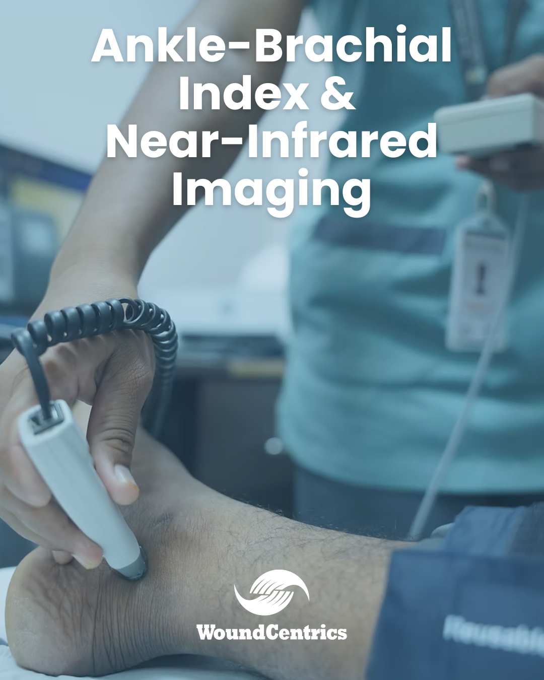
One of the reasons for this is that commonly available vascular screening methodologies, such as Ankle Brachial Index, and the related Toe Brachial Index suffer from significant limitations in the diabetic populations common in our wound centers. Factors such as vascular calcification which are common in these patients render ABI data difficult to interpret. TBI addresses some of these limitations, but it can be very difficult to reliably administer the test in a significant percentage of patients.

More importantly, ABI and TBI do not tell us anything about microvascular flow in the wound bed, and certainly nothing about tissue oxygenation – an essential factor for wound healing.

Transcutaneous Oxygen Mapping, or TCOM, long accepted as the “Gold Standard” for assessing tissue oxygenation (and collaterally, microvascular blood flow) and thereby assessing potential for wound healing, also suffers from very significant limitations in clinical practice.

One key limitation of TCOM is that it cannot be used to directly assess perfusion of plantar skin; the site of some of our most challenging wounds. In addition, the TCOM electrodes in common use are not suitable for assessing perfusion in the toe, due to their size.

In addition to these technical limitations, TCOM is an expensive technology to purchase and maintain, with typical devices costing more than $50,000 to purchase. My group recently received a $76,000 quote for maintenance costs for six devices. They also require a significant training investment to use effectively, and testing can be time consuming, consuming valuable technician time and bed minutes in the clinic.

In an era of decreasing margins, and increasingly stringent demands for efficient, optimal utilization, the limitations and expenses associated with ABI and TCOM are forcing clinical leaders, and clinic owners to look to new technologies offering a better cost-benefit ratio.

As I pointed out in my recent lecture, two technologies are generating enthusiasm for new paradigms in non-invasive vascular evaluation in the wound clinic and bringing hope that wound care programs can grow beyond the current technical limitations and cost burdens imposed by existing technologies. The two technologies I focused on are Combined Skin Perfusion Pressure and ABI with PVR (Vasomed PAD-IQ), and Hyperspectral Imaging (Kent Imaging “Snapsho2t,” and Hypermed “Hyperview”).

While Hyperspectral Imaging and Skin Perfusion Pressure are very different from a technological perspective, each of these new devices address the imperative to quickly, repeatedly, and cost-effectively assess the vascular status of our patients, so that treatment decisions can be optimized from the point of care, on the first visit.

Hyperspectral imaging technology is not fundamentally new, but the use of hyperspectral imaging to assess skin blood flow is a recent development. Many wound care clinicians have seen one or more of these devices demonstrated at industry trade shows, such as SAWC, and those who have seen them are typically intrigued by what they see.

These units are just a bit bigger than an iPAD, and are capable of capturing an image that provides direct, quantitative visualization of oxygenated hemoglobin, deoxygenated hemoglobin, and oxygen saturation, superimposed on a visual image of the wound and surrounding skin.

Hyperspectral images can be obtained in seconds, without expendable costs, by a technician or provider with as little as five minutes of training, and they are compelling to look at, but are they useful clinically? The literature is meager right now, but several groups are publishing clinically oriented papers assessing the utility of the technology for wound care and vascular medicine.

These studies have looked at the predictive value of hyperspectral imaging in patients with vascular ulcers and diabetic foot ulcers, and what has been consistently evident is that deoxy-hemoglobin imaging data correlates reasonably well with TcPO2:

TcPO2 and DeOxyHgb (r2 = 0.63, P < 0.0001)2

Hyperspectral imaging also correlates with angiosomal anatomy:

“Deoxyhemoglobin values for the plantar metatarsal, arch, and heel angiosomes were significantly different between patients with and without PAD (P <.005)”3

Finally, and perhaps most meaningfully, in a study of 73 diabetic foot ulcers in 66 patients, over a 24 week period, hyperspectral imaging correctly predicted healing with 80% sensitivity, and non-healing with 74% Specificity.4 Thus as a predictor of healing, hyperspectral imaging compares quite favorably with TCOM, whose generally accepted sensitivity for prediction of healing is 72%.

Having evaluated one of these units in a wound clinic setting, I find the information helpful, but difficult to integrate into the practice patterns necessitated by LCD strictures, and commonly accepted treatment pathways. I intend to watch these devices evolve, and champion their promise. They likely represent a future evolutionary step in wound care and vascular medicine, but I don’t think they are a compelling replacement for TCOM at this time.

If Hyperspectral Imaging is not ready for prime time, Skin Perfusion Pressure may be ready to step into the breach and meet the need for timely and cost-effective evaluation of tissue perfusion in the critical DFU and vascular ulcer population. While not a new technology, SPP is now available in the form of an integrated, elegant device called PAD-IQ, capable of providing rapid, repeatable, predictive test results in the challenging lower extremity wound population.

By combining ABI testing with Pulse Volume Recording (PVR), PAD-IQ can deliver 100 sensitivity for identification of PAD, and 100% negative predictive value for absence of the disease. Integrative SPP allows clinicians to perform accurate and repeatable “perfusion maps,” much like those obtained using multi-channel TCOM testing, but unlike TCOM, users can assess perfusion at the toe, as well as plantar skin.

Having used one of these devices in the wound clinic for 6 weeks, I can say unequivocally that I am ready to give up my TCOM device and embrace a new way of assessing skin perfusion. The PAD-IQ is a well-designed device that meets multiple needs in one unit. Nurses and technicians were quickly trained and performing studies. The perfusion sensor is easy to apply, and the interface is about as elegant as any I have seen on a medical device. Printouts are easy to read, and information rich; the kind of reports one is proud to send to a referral source, or consultant. Results are unambiguous, and graphically clean.

Compared to TCOM, a PAD-IQ test can be completed in less than half the time. Like TCOM, interpretation of SPP is straightforward. Using a cutoff of 30mmHg (capillary opening pressure, not TcPO2), one can readily predict non-healing status, and determine the need for intervention.

How does SPP compare to TCOM in clinical trials? Lo, et. al. found that SPP alone successfully predicted wound outcome in 87% of the cohort compared to TcPO2 at a rate of 64% (P < 0.0002). In addition, SPP was more sensitive in predicting wound healing than TcPO2 (90% versus 66%; P <0.0001).5

These are impressive results, and I feel just what we need to move the management of lower extremity ulcers, and particularly diabetic foot ulcers forward. Too many programs are confounded by unsystematic, subjective vascular evaluation heuristics.

It is as though we have been evaluating vascular status in these technically complex, challenging cases using a “magic 8 ball.” I believe that, until we are assessing every lower extremity wound patient with PAD risk factors early, and definitively, we will be “running in place” as a specialty, generating inconsistent, suboptimal outcomes.

It is time, I feel, to make vascular testing simple, reliable, and something that happens at the bedside at the point of consultation in the wound clinic. I for one am ready to embrace change. Please let me know your thoughts and insights. I’ll be presenting on this topic next year at ACHM, where I look forward to sharing much more data.

Marcus Gitterle, M.D., FACCWS
Chief Medical Officer
WoundCentrics, LLC


__________________________________________
1Centers for Disease Control, 2019
2Jafari-Saraf L, Wilson SE, Gordon IL. Hyperspectral image measurements of skin hemoglobin compared with transcutaneous PO2 measurements. Annals of Vascular Surgery. 2012;26(4):537–548
3Chin JA, Wang EC, Kibbe MR. Evaluation of hyperspectral technology for assessing the presence and severity of peripheral artery disease. Journal of Vascular Surgery. 2011;54(6):1679–1688
4Bolton, L., Hyperspectral Imaging: Early Warning of Low Tissue Perfusion Wounds, 2012;24(10):A8-A105 Lo, T, et al, Prediction of wound healing outcome using skin perfusion pressure and transcutaneous oximetry: a single center experience in 100 patients, Wounds, 2009 Nov: 21(11) 310-6
5Lo, T, et al, Prediction of wound healing outcome using skin perfusion pressure and transcutaneous oximetry: a single center experience in 100 patients, Wounds, 2009 Nov: 21(11) 310-6

A Message from WoundCentrics, NALTH Visionary Partner: The Evolving Role of Wound Care in Long Term Acute Care

The Evolving Role of Wound Care in Long Term Acute Care

With the FY 2020 phase out of blended payments beginning October 1, 2019, most LTACHs have begun severely limiting, or eliminating altogether, non-qualifying LTACH admissions. As expected, this has greatly reduced the number of wound related DRGs admitted and discharged from long term acute care hospitals. As these patients are more and more often cared for in other settings one might reasonably expect that the importance of wound care in the LTACH would be decreased. While some facilities diversify their scope of service to include co-located skilled nursing facilities or inpatient rehab beds, others have developed outpatient wound centers to meet patient needs.

As a company devoted exclusively to wound care, across the continuum of care, it is obvious to us that the severe wounds we formerly cared for are no longer commonly seen as the primary reason for admission in the LTACH. We do care for them in many other levels of care that we previously did not. However, rather than the loss of primary wound care admissions resulting in a decrease in the incidence of wounds in the LTACH setting, the new LTACH admission criteria appears to have resulted in an equal and often increased incidence of wounds in the nearly two dozen LTACHs WoundCentrics serves. In addition, in the dozens of acute care hospitals we provide care in, we see qualifying patients with wounds go unreferred or unaccepted by our LTACH partners.

Even in those LTACHs where the incidence of wounds has decreased, the DRG case weight changes almost always result in equivalent or greater financial returns in response to wound care than were previously seen with DRG changes related to patients with primary wound care related DRGs. Consider the two examples below;

A 592 DRG (SKIN ULCERS WITH MCC) HAS A CASE WEIGHT OF 0.9629 AND IF DEBRIDED WOULD BECOME A DRG 570 (SKIN DEBRIDEMENT WITH MCC) WITH A CASE WEIGHT OF 1.2916. WITH THE LTACH-PPS STANDARD RATE OF $42,677.64 THAT WOULD INCREASE THE DRG PAYMENT BY $14,028.34

A DRG 207 (RESPIRATORY DIAGNOSES WITH >96 HOURS VENTILATOR SUPPORT) AND A CASE WEIGHT OF 1.8628 WOULD BECOME A DRG 166 (OTHER RESPIRATORY SYSTEM OR PROCEDURE WITH MCC) IN MOST CASES AND CARRY A CASE WEIGHT OF 2.3392. THE RESULTING DRG PAYMENT WOULD INCREASE BY $20,331.27. PATIENTS WHO DID NOT MEET THE 96 HOURS OF VENTILATOR SUPPORT SUCH AS DRG 208 ($47,966.25 INCREASE) OR DRG 189 ($58,792.72 INCREASE) RESULT IN AN EVEN GREATER IMPACT


As you can see, wound care still has an important role to play in the LTACH.

We invite you to stop by the WoundCentrics booth while at the Fall NALTH Conference or grab one of our team members any time during the conference and let us give you some examples of the real life experiences our LTACH partners have seen in their facilities this year.

Company Contact
Ken Rideout, Development
kenrideout@woundcentrics.com
Cell / Text: 281-989-5398

Stuart Oertli, COO, Asks: “Are you Planning for the New LTACH Rules this Year?” What are your plans to combat a decrease in admissions and lost revenue?

The WoundCentrics Specialized Wound Services program, designed for Long Term Acute Care Hospitals, is proving to be an option to help hospitals flourish under the new LTACH rules.  Our focus on wound care services will improve quality wound care, improve clinical alignment for both patients and providers all while continuing to demonstrate a substantial return on investment for hospitals who contract with WoundCentrics.  

I would like to offer a couple of observations from our newest LTAC Hospital partner.  Prior to engaging WoundCentrics,  there was significant concern from both local administration and corporate leadership that the need for wound care would be greatly diminished as blended payments phase out starting this fall.  Additionally, there was no plan to admit primary wound DRG patients once true site neutral reimbursement began.

The argument against incurring a new expense for a specialized wound care program was that the LTAC Hospital currently had a very well respected, albeit a very busy, local wound care Medical Director in place.   He was affiliated with multiple Acute Care Hospitals and usually, rounded at the LTACH once or twice a week, typically at the end of the day or after 6pm.  Due to his busy day schedule, it was impossible for him to fully integrate with the hospital’s bi-weekly IDT team meetings, nor did he have the time to support the facility wound care nurse on a daily basis.

Additionally, the LTACH facility was already staffed with a well educated, employed wound care team already in place.  The facility had a strong corporate commitment to quality and decision support to assist the staff on the ground.  The local CEO was recently recruited from a major LTAC Hospital ownership group, with a great deal of LTACH experience and pushed to implement our Specialized Wound Services program and worked hard to receive the support from his corporate leadership and full buy-in from his clinical team.

Here is the ROI example I wanted to share: 

The WoundCentrics employed Nurse Practioner wound specialist started seeing patients under the management of the WoundCentrics program at this 38 Bed LTACH in early May.  During the first 2 weeks,  the clinical team identified and completed 7 unique wound debridements,4 of them on LTACH compliant admissions despite the fact that their current payer mix was about 50% site neutral, 50% LTACH compliant). 1 of the debridements was on a patient with a commercial payer who extended authorization due to the active wound care being provided and the final 2 were site neutral Medicare payers.

Four (4) of the wound debridements have been coded thus far and are shown below. I provide this information in hopes that ROI will be immediately evident: 

Patient 1: Admission DRG 207 (1.8542), Post-Debridement DRG 166 (2.4628). Revenue Increase $27,223.83 

Patient 2: Admission DRG 592 (0.9330), Post-Debridement DRG 570 (1.3477). Revenue Increase $18,550.32 

Patient 3: Admission DRG 207 (1.8542), Post-Debridement DRG 166 (2.4628). Revenue Increase $27,223.83 

Patient 4: Admission DRG 208 (1.1033), Post-Debridement DRG 166 (2.4628). Revenue Increase $60,813.02 

Total Revenue Increase from first two weeks of program = $133,811.00.

Assuming net of 50% after expenses (incremental costs are likely much less), the net revenue would be $66,805.50. We proposed a $7,500 monthly fee for single locations, and a corporate rate is available for multiple facilities.  Thus, the entire first year program cost was covered within 2 weeks of program implementation and before considering the additional benefits from commercial and site neutral revenue changes. 

During the third week, 2 additional unique debridements were completed bringing the total revenue increase from the first three weeks of the program to $195,975.29:

Patient 5: Admission DRG 699 (0.6746), Post-Debridement DRG 856 (1.5413). Revenue Increase $34,940.46

Patient 6: Admission DRG 207 (1.8542), Post-Debridement DRG 166 (2.4628). Revenue Increase $27,223.83

This early success with our Wound Services program has our new LTACH client off to a great start.  Continued focus on quality wound care and constant revenue cycle review with administration will provide the revenue to keep the facility financially viable and in position to offer their valuable services to patients in their healthcare community.

What is Your Plan to Survive the new LTACH Rules?

The WoundCentrics Specialized Wound Services program has provided quality wound care for all our facilities and the revenue example provided above repeatable and reliable. It is consistent with every LTACH facility where we have been able to implement our full program with the support of and in coordination with the locate wound care team.  We have historical performance data that shows we have achieved similar results (many times far better) in every single location where we've created an LTACH partnership.

If you are considering plans to attack lost revenue related to decreased volumes and full implementation of site neutral payments for LTAC Hospitals in the coming months, please contact us for more information on how the WoundCentrics Specialized Wound Services program might work with the Wound Care team in your facility. 

For more details about WoundCentrics visit our web site:    www.woundcentrics.com                                                              Contact:  Ken Rideout, VP Development(281) 989-5398

Wound research collaboration

Though woundcare is now a well established specialty, fundamental questions remain unanswered with respect to the pathologies which result in non-healing wounds. WoundCentrics is helping to champion the cause of basic wound research by promoting provider involvement in active research.

Case in point is an exciting partnership between the US Army Institute for Surgical Research (ISR), and one of our clinicians, CEO Marcus Gitterle, MD. The ISR was founded to focus on the scientific aspects of wound healing, an issue that impacts countless warfighters and civilians annually. The capabilities of their primary lab in San Antonio, Texas, are without peer in this field.

To help advance the clinical science of wound healing, Dr. Gitterle began a long-term collaboration with the US Army, designed to harness the synergy between our large clinical footprint and provider expertise, and their best-of-breed laboratory expertise.

The first study under this collaborative agreement was launched in February, 2016. This study is expected to be the first complete study of the human wound microbiome, including all bacterial, fungal and viral components.

In addition, the study represents the first use of “transcriptomics,” in non-healing wounds, allowing researchers to analyze signaling between microbiome organisms, thought to be a potential source for healing interventions.

Our goal in this research collaboration is to help advance understanding of the complex role of the human microbiome in contributing to non-healing wounds, and ultimately to help identify solutions to wound healing challenges through this understanding.

We are aware that many organizations in woundcare pursue research affiliations, but these are predominantly relationships that create revenue, and the focus is predominantly on products, rather than fundamental scientific insight.

WoundCentrics is proud to facilitate important research that is not funded by industry, and which has the potential to significantly advance the science of wound healing.  It’s our way of “giving back.”